

Biological Science Research Facilities
The Biological Imaging Facilities (BIF) are available to graduate students, postdoc researchers, and faculty affiliated with UAlbany, as well as academic and non-profit researchers in the Capital Region.
Interested users are invited to contact the BIF Director using their official institutional email. Please include the supervising Principal Investigator who will serve as the billing contact for hourly service charges.
Facilities
Confocal Laser-Scanning Microscopy
Confocal Laser-Scanning Microscopy
Zeiss LSM 980
The Zeiss LSM 980 system is the up-to-date confocal laser scanning microscope with Airyscan 2 detection. The major technical specifications are outlined as follows:
LSM 980 Stand
- Inverted motorized Axio Observer 7 SP with Definite Focus 3 nosepiece
- Motorized XY Stage with inserts for slides, well-plates, petri-dishes, and chambered slides
LED excitation sources (Colibri 5) for wide-field fluorescence imaging by eyepiece observation
Color Wavelength (nm) Related filter Cube UV 385 DAPI Blue 475 GFP Green 555 RFP Red 630 Cy5 - Motorized condenser with HAL 100 for bright-field imaging by eyepiece observation
- Transmission PMT (T-PMT) detection for DIC imaging by transmitted laser beam & motorized condenser lens & polarizer and prism & computer monitor display
- Active anti-vibration table to facilitate fluorescence-bead-like imaging
LSM 980 Excitation Laser
| Laser Type | Laser Wavelength (nm) | Minimal acceptable power ex-fiber (mW) |
|---|---|---|
| Diode | 405 | 11.3 |
| Diode | 445 | 18.5 |
| Diode | 488 | 16.9 |
| Diode | 514 | 12.1 |
| DPSS | 561 | 14.3 |
| DPSS | 594 | 4.8 |
| Diode | 639 | 12.7 |
| Diode | 730 | 9.8 |
LSM 980 Objective
| Type | Magnification | NA (numerical aperture) | Free working distance (mm) | Coverglass thickness (mm) | Immersion type |
|---|---|---|---|---|---|
| EC Plan-Neofluar | 10x | 0.3 | 5.2 | 0.17 | Air |
| Plan-Apochromatic | 20x | 0.8 | 0.55 | 0.17 | Air |
| Plan-Apochromatic | 40x | 1.4 | 0.13 | 0.17 | Oil (Immersol 518F) |
| Plan-Apochromatic | 63x | 1.4 | 0.19 | 0.17 | Oil (Immersol 518F) |
- Touchscreen for objective selection
LSM 980 Confocal Laser Scanning Microscopy (cLSM) mode
- Simultaneous multi-color (up-to 4) imaging & sequential multi-color (up-to 12) imaging by Smart Setup imaging scheme with 34-channel PMT-array spectral detection
- Sequential single-color imaging with no spectral cross-talk by lambda & sum-up multi-color imaging scheme with 34-channel PMT-array spectral detection
- Expanded fluorescence imaging to near-infrared range with Diode laser excitation @730nm & near-infrared detector pair in addition to 34-channel PMT-array visible spectral detection
LSM 980 Airyscan 2 Imaging mode
- Imaging software menu preset excitation & detection proposals in pursuit of ultra-high image resolution, ultra-fast image acquisition, or ultra-high sensitivity of dim-signal detection
- Imaging software menu preset excitation & detection proposals for customized image acquisitions in terms of image resolution or acquisition speed
LSM 980 Cooling Unit
- Excellent signal-to-noise ratio detection of fluorescence signals achievable with 34-channel PMT-array visible spectral detection, near-infrared spectral detection, and Airyscan 2 detection owing to distilled-water circulation cooling system
LSM 980 Z-Stack Imaging mode
- 3D imaging with thick samples to generate image cube from 2D slices acquired by either cLSM or Airyscan 2 imaging schemes
- 3D imaging with thick samples to generate larger FOV (field-of-view) image cube by the above-mention 3D imaging & tile imaging
LSM 980 Incubation Chamber (transparent)
- Gas mixer system with CO2-control range 0 – 15%; Humidity control range 20 – 99%
- Heating environment for Chamber, well-plate, or petri-dishes
Zeiss LSM 710
The Zeiss LSM 710 system’s major technical specifications are outlined as follows:
LSM 710 Stand:
- Inverted Axio Observer.Z1with Definite Focus 3 nosepiece
- Motorized XY Stage with inserts for slides, well-plates, petri-dishes, and chambered slides
- X-Cite 120PC Q lamp for wide-field fluorescence imaging by eyepiece observation
- Motorized condenser with light source of HAL 100 for bright-field imaging by ocular observation
- Transmission PMT (T-PMT) detection for DIC imaging by transmitted laser beam & motorized condenser lens & polarizer and prism & computer monitor display
- Active anti-vibration table to facilitate fluorescence-bead-like imaging
LSM 710 Excitation Laser:
| Laser Type | Laser Wavelength (nm) | Minimal acceptable power ex-fiber (mW) |
|---|---|---|
| Diode | 405 | 30 |
| Argon | 458,488,514 | 25 |
| DPSS | 561 | 20 |
| He-Ne | 633 | 5 |
LSM 710 Objective
| Type | Magnification | NA (numerical aperture) | Free working distance (mm) | Coverglass thickness (mm) | Immersion type |
|---|---|---|---|---|---|
| Plan-Apochromatic | 10x | 0.45 | 2.0 | 0.17 | Air |
| Plan-Apochromatic | 20x | 0.8 | 0.55 | 0.17 | Air |
| EC Plan-Neofluar | 40x | 1.3 | 0.21 | 0.17 | Oil (Immersol 518F) |
| Plan-Apochromatic | 63x | 1.4 | 0.19 | 0.17 | Oil (Immersol 518F) |
- Touchscreen for objective selection
LSM Confocal Laser Scanning Microscopy (cLSM) mode:
- Simultaneous multi-color (up-to 4) imaging & sequential multi-color (up-to 12) imaging by Smart Setup imaging scheme with 34-channel PMT-array spectral detection
- Sequential single-color imaging with no spectral cross-talk by lambda & sum-up multi-color imaging scheme with 34-channel PMT-array spectral detection
LSM 710 Z-Stack Imaging mode:
- 3D imaging with thick samples to generate image cube from 2D slices acquired by either cLSM mode
- 3D imaging with thick samples to generate larger FOV (field-of-view) image cube by the above-mention 3D imaging & tile imaging
LSM 710 Incubation Chamber (transparent):
- Gas mixer system with CO2-control range 0 – 15%; Humidity control range 20 – 99%
- Heating environment for Chamber, well-plate, or petri-dishes
Digital Fluorescence Microscopy
Digital Fluorescence Microscopy
EVIDENT VS200 Slide Scanner & Loader System


Illumination Sources:
X-Cite NOVEM-L, XT920 w/1.5m LLG 380-770 nm
FLUOROPHORE | WAVELENGTH (nm) |
DAPI | 385 |
CFP | 435 |
FITC | 475 |
YFP | 510 |
TRITC | 555 |
mCherry | 580 |
CY5 | 635 |
CY7 | 735 |
Slide Dimension:
1”x3” compatible with current trays
Objectives:
Type | Magnification | NA (numerical aperture) | Free working distance (mm) | Cover glass thickness (mm) | Immersion type |
ACH | 2x | 0.06 | 5.8 | 0.17 | Air |
UPLAXPO | 4x | 0.16 | 13 | 0.17 | Air |
UPLAXPO | 20x | 0.8 | 0.6 | 0.17 | Air |
UPLAXPO | 40x | 0.95 | 0.18 | 0.17 | Air |
UPLAXPO | 40x | 1.4 | 0.13 | 0.17 | Oil |
UPLAXPO | 60x | 1.42 | 0.15 | 0.17 | Oil |
Image Acquisition:
ORCA-Fusion Gen-III sCMOS CAMERA. C-mount with USB 3.0 card (capable of full-frame acquisition at a rate up to 30 frames per second (fps). VS200 Acquisition Software (ASW) + Batch File Conversion Module
2D Imaging Modes:
Bright-field imaging
Darkfield imaging
Fluorescence imaging with custom Penta emission filters for DAPI, CFP/YFP, FITC, mCherry, TRITC, Cy5 and Cy7, applicable to other fluorophores of similar emission wavelengths
Microscopy Image Analysis Workstation
Microscopy Image Analysis Workstation
Microscopy Image Analysis Software
- Imaris (OXFORD INSTRUMENT)
- ImageJ (NIH)
- MetaMorph (MOLECULAR DEVICES) for 3D deconvolution or reconstruction; particle tracking & motion analysis; FRET, FRAP, and FISH image analysis; multispectral image segmentation; and time-lapse image analysis
- MetaFlour (MOLECULAR DEVICES) for fluorescence ratio (Calcium, FRET, or FLIM) image analysis
Two-Photon Laser Scanning Microscopy
Two-Photon Laser Scanning Microscopy
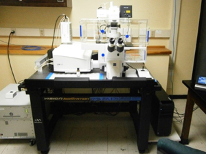
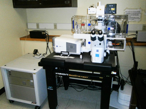
A custom-made two-photon laser scanning microscope is available, being compatible to electrophysiological measurement facilities for research in neuroscience.
Interested parties may contact Dr. Scimemi for details.
Facility Usage Fees & Rules of Reservation
Facility Usage Fees & Rules of Reservation
Usage Fee Policy
- Usage Fee Policy
Instrument | User Type | Approved Users | Assisted Users | Training session | Image Analysis Workstation |
Zeiss LSM980 | UAlbany Users1 | $25/hour | $50/hour | $50/hour | N/A |
Off-campus | $35/hour | $60/hour | $60/hour | $10/hour | |
| Users2 |
|
|
|
|
Evident VS200 | UAlbany Users1 | $10/hour | $20/hour | $30/hour | N/A |
Off-campus | $25/hour | $50/hour | $60/hour | $10/hour | |
| Users2 |
|
|
|
|
- Graduate students, Postdoc researchers, and Faculty affiliated with UAlbany.
Academic & Non-profit researchers in the Capital Region.
Rules of Reservation
Each approved user can reserve the usage of an instrument for up to 3 hours during 9 AM – 5 PM on a weekday. More-than-3-hour reservations are encouraged outside business hours & on weekends.
Donations
Donations
Our facilities cannot function without the generous support from a variety of sources
These sources include:
- Shared Equipment grants from the National Science Foundation.
- Support from the College of Arts & Sciences and from the Research Office of SUNY.
- Fees paid by our users.
- Financial Support from individuals.
Donations for operations or expansion of the Imaging Center can be made by contacting the Department of Biological Sciences.
Please contact:
- The Biology Department Chair, Dr. Melinda Larsen
- The University's Development Officer, Michael Boots
- The Division of University Advancement
Contact Information
Contact Dr. Chao Wang, Director of Biological Imaging Facilities (BIF), for detailed information.