

The Tissue Culture Core Facility is a BSL 2 common use facility designed for the culturing and manipulation of primary tissue and mammalian cell lines. It provides a variety of instrumentation that facilitates research in Virology, Developmental Biology, Physiology, Immunology, Neurobiology, Cancer, and Behavioral Science that requires the cultivation of cell lines or tissues. The Tissue Core Facility provides maintenance, scheduling and training on instrumentation and University at Albany investigators can access the facility anytime. Essential equipment used to prepare, manipulate, image, incubate, harvest and store samples are provided in support of investigators’ research.
Equipment
For access or consultation, contact Heather Edgerton at 518-591-8831 or [email protected].
Scroll this page and learn more about our equipment use by clicking on the links below:
Microscopes
Nikon Eclipse TS100 Inverted Microscopes (3)
Nikon Eclipse TS100 Inverted Microscopes (3)
The Tissue Core Facility has three Nikon Eclipse TS100 inverted microscopes.
Purpose
Microscopes are used to visualize, analyze, document and manipulate cells at magnifications much greater than that observed under normal vision.
Specific Description
The TS100 inverted microscopes are equipped with 10x eyepieces and 10x, 20x and 40x objectives.
More Information
Nikon Eclipse TS100 Manual
Leica MZ16FA fluorescent dissecting microscope
Leica MZ16FA fluorescent dissecting microscope
The Tissue Core Facility has one Leica MZ16FA fluorescent dissecting microscope.
Purpose
Microscopes are used to visualize, analyze, document and manipulate cells at magnifications much greater than that observed under normal vision. Fluorescent microscopes allow researchers to identify specific cell types and locations of cells labeled with specific fluorophores.
Specific Description
The LeicaMZ16FA has a 10x eyepiece and 1x objective with a motorized zoom range of 7.1x to 115x. Images can be seen through the eyepiece or directed to a computer monitor. There are three fluorescent filter sets used with the excitation/emission wavelengths 360 nm/420 nm, 480 nm/510 nm and 545 nm/620 nm.
More Information
Leica MZ16FA User Manual
Evos Fl Cell Imaging Microscope
Evos Fl Cell Imaging Microscope
The Tissue Core Facility has one Evos Fl Cell Imaging Microscope
Purpose
Microscopes are used to visualize, analyze, document and manipulate cells at magnifications much greater than that observed under normal vision. Fluorescent microscopes allow researchers to identify specific cell types and locations of cells labeled with specific fluorophores.
Specific Description
The Evos FL cell imaging microscope uses either transmitted light or fluorescence to visualize live or fixed cells in culture plates, 96 well plates or slides. It is equipped with 2x, 4x, 10x, 20x, and 40x objectives. There are six fluorescent filter sets used with the excitation/emission wavelengths 357 nm/447 nm, 445 nm/510 nm, 470 nm/510 nm, 500 nm/524 nm, 531 nm/593 nm, 585 nm/624 nm and 628 nm/692 nm.
More Information
EVOS FL Manual
Centrifuges
Beckman-Coulter Centrifuges (2)
Beckman-Coulter Centrifuges (2)
The Tissue Culture Core Facility has a Beckman-Coulter Avanti J-E Centrifuge and an Avanti J25 Centrifuge.
Purpose
Centrifugation is a process where samples are spun at very high speeds to create gravitational forces (g force) great enough to precipitate cells, organelles and insoluble components in a mixture.
Specific Description
These floor model centrifuges are used for medium speed centrifugation. Volumes from less than 10 milliliters up to 3 liters can be prepared in these centrifuges depending on the rotor used.
Available Rotors in the Tissue Culture Core Facility
|
Rotor |
Max (rpm) |
Max (g force) |
Tube/bottle size |
Capacity |
|---|---|---|---|---|
|
Fixed Angle Rotors |
||||
|
JA-10 |
10,000 |
17,000 |
250 ml |
6 |
|
JA-12 |
12,000 |
17,400 |
50 ml |
12 |
|
JA-14 |
14,000 |
18,900 |
250 ml |
6 |
|
JA-20 |
20,000 |
31,400 |
50 ml |
8 |
|
JA-20.1 |
20,000 |
47,900 |
15 ml |
32 |
|
JA-25.5 |
25,000 |
51,200 |
50 ml |
8 |
|
Swinging Bucket Rotors |
||||
|
JS-13.1 |
13,000 |
17,200 |
50 ml |
6 |
More Information
Avanti JE Operation Manual
Eppendorf 5810 & 5810R Clinical Centrifuges
Eppendorf 5810 & 5810R Clinical Centrifuges
The Tissue Culture Core Facility has one refrigerated clinical centrifuges (Eppendorf 5810R) and one clinical centrifuge without refrigeration (5810).
Purpose
Clinical centrifuges are used for a variety of separations, but primarily to pellet cells from medium using relatively low g forces (3220 x g). Another common use is to spin membrane devices that separate macromolecules based on size.
Specific Description
These centrifuges are equipped with swinging bucket holders that accommodate either 4 round or 4 rectangular shaped holders. The rectangular shaped holders can be used to spin multi-well plates, 15 ml culture tubes or 50 ml culture tubes. The round holders are used to spin either 15 ml culture tubes or 50 ml culture tubes. Each rectangular holder can support three 50 ml tubes, nine 15 ml tubes or one multi-well plate. Each round holder can support seven 50 ml tubes or fourteen 15 ml tubes.
More Information
Eppendorf 5810 Centrifuge Manual
Eppendorf 5415D Centrifuges (6)
Eppendorf 5415D Centrifuges (6)
The Tissue Culture Core Facility has six microfuges without refrigeration (Eppendorf 5415D).
Purpose
A microfuge is a workhorse type of centrifuge used for a variety of separations in small volumes typical of Molecular Biology. The maximum centrifugal force in these centrifuges is 16,000 x g.
Specific Description:
These centrifuges are equipped with fixed angle rotors that accommodate 24 in non-refrigerated microfuges. The rotors can hold either 2.0 ml vials, 1.5 ml vials, 0.5 ml vials or 0.2 ml vials with adapters.
More Information
Eppendorf 5415D Centrifuge Manual
Incubators
New Brunswick Incubator/Shakers
New Brunswick Incubator/Shakers
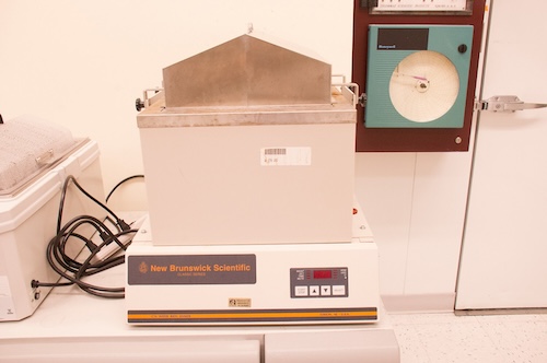
The Tissue Core has one New Brunswick C76 water incubator/shaker.
Purpose
Water baths are typically used to heat medium prior to inoculation with tissue culture cells.
Specific Description
The New Brunswick C76 water bath heats from 7°C over ambient up to 80°C and can shake from 50 to 300 rpm.
For More Information
NB C76 Water Incubator Shaker Manual
Forma 3110 CO2 Incubators (4)
Forma 3110 CO2 Incubators (4)
The Tissue Core has four Forma 3110 CO2 incubators.
Purpose
CO2 incubators are typically used to maintain a 37°C temperature and 5% CO2 gas phase for tissue culture cells in liquid or solid medium.
Specific Description
The Forma 3110 CO2 incubator heats from 5°C over ambient up to 55°C, with up to 95% humidity at 37°C and CO2 concentration from 0 to 20%.
More Information
Forma 3110 CO2 Incubator Manual
New Brunswick 170S CO2 Incubators (2)
New Brunswick 170S CO2 Incubators (2)
The Tissue Core has two New Brunswick Galaxy 170S CO2 incubators.
Purpose
CO2 incubators are typically used to maintain a 37°C temperature and 5% CO2 gas phase for tissue culture cells in liquid or solid medium.
Specific Description
The New Brunswick 170S CO2 incubators heats from 4°C over ambient up to 50°C, with up to 95% humidity at 37°C and CO2 concentration from 0.2 to 20%.
For More Information
Galaxy S Series 170S CO2 Incubator Manual
Water Purification
Water Purification System
Water Purification System
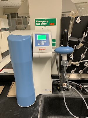
The Tissue Culture Core Facility has a Barnstead GenPure Pro Ultrapure water system.
Purpose
Using water purified of biological, organic and inorganic contaminants is essential for the preparation of buffers, medium and reagents used in culturing cells, enzymatic or chemical assays and analytical procedures.
Specific DescriptionThe Barnstead GenPure Pro Ultrapure water system is connected to the buildings reverse osmosis purified water source and uses a four-stage deionization process combined with a UV lamp, an ultrafilter and a 0.2 micron filter to polish feed water to produce pyrogen-free, low TOC water with a resistivity of up to 18.2 MΩ-cm.
For More Information
Barnstead GenPure Pro Ultrapure Water System Manual
Storage and Preservation
Freezers and Refrigerators
Freezers and Refrigerators
The Tissue Culture Core Facility has a New Brunswick U700 -80°C freezer, two Taylor Wharton LS6000 liquid storage tanks with an autotend feature and two Labconco Freezone 4.5 lyophilizers.
Purpose
Many biological samples including cells, reactions, tissues, reagents, etc. require storage at temperatures at 4°C, -25°C, -80°C or -195°C. Other biological materials are freeze dried in lyophilizers to remove water so as to concentrate them, limit their decomposition and facilitate their storage.
Specific Description
Each -80°C freezer has two compressors to achieve the low temperatures, a side vent to release vacuum before opening and an alarm system that sounds when the freezer’s temperature is above -70°C. The Taylor Wharton LS 6000 cryogenic storage tanks are filled with liquid nitrogen via an electronic monitoring device called autotend. The autotend allows liquid nitrogen to fill the tank when it falls below the lower sensor and fills it up to the upper sensor. The Labconco lyophilizers have refrigeration units that achieve temperatures below -40°C and are connected to an Edwards XDS 15 oil-less scroll pump to achieve vacuum levels below 0.1 mBar.
More Information
Labconco Freezone Manual
Taylor Wharton LS6000 liquid storage tank Manual
Sample Preparation
Sample Preparation Equipment/Instruments
Sample Preparation Equipment/Instruments
The Tissue Culture Core Facility also has an Mettler Toledo AB204-S/Fact analytical balance, Denver Instruments APX-153 top loading balance, Mettler Toledo Seven easy pH meter, Barnstead Diamond Nanopure water purifier and a Consolidated Sterilization Systems autoclave. The Tissue Core has four Baker Company Sterilgard III Advance laminar flow hoods, a Beckman-Coulter Z2 coulter particle count and size analyzer, a Leica CM 1860 cryostat, a Leica VT 1200S vibratome and a Leica RM 2255 Microtome.
Purpose
Sample preparation is a critical component to any experiment. Basic procedures like using nanopure water, weighing chemicals, measuring pH of buffers and medium and sterilizing glassware, utensils, buffer are essential to all experiments. Sample preparation includes manipulating cells, tissues or biological materials in an appropriate environment and making thin sections for microscopic investigation. The Tissue Core facility has instruments that are designed to facilitate sample preparation. The particle counter can count the number of cells, give the concentration and the size distribution of a cell population.
Specific Description
Each laminar flow hood is designed to direct airflow around a surface where cell and tissue manipulation occurs to minimize contamination. The atmosphere is continuously filtered with HEPA filters and ultraviolet light illumination keeps the surface sterile while not in use.
The vibratome is used to prepare histology samples for microscopic analysis. The speed that the vibratome sections material can be adjusted from 0.01 to 1.5 mm/s and the amplitude of sections can be adjusted from 0 to 3 mm in increments of 0.05 mm.
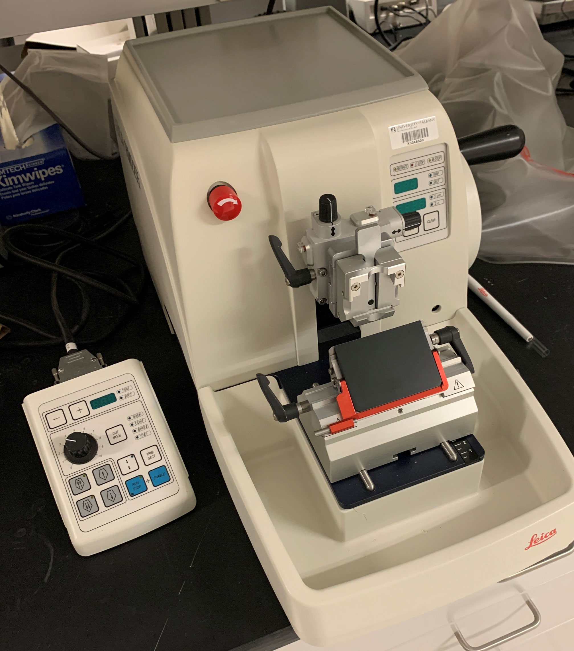
The Microtome is used to prepare parafin imbedded tissues for microscopic analysis. The parafin and tissue are sliced into 0.5 mm to 100 mm thick sections. The parafin is melted at 55°C leaving the tissue section for analysis.
The cryostat is used to prepare frozen tissues for microscopic observation. The cryostat chamber can be set from 0°C to -35°C and the section thickness can be adjusted from 1 to 100 μm.
The particle counter uses the electrical sensing zone method of counting and sizing of cells thus providing accuracy, speed, versatility and reproducibility. The counter provides the entire size distribution graph, the cumulative count and the ability to average count data from a series of up to 10 consecutive analyses.
More Information
Leica Cryostat Manual
Leica Vibratome Manual
Microtome Manual
Beckman-Coulter Z2 Particle Counter Manual
Sterilgard III Advance Hood Manual
Other Equipment
Cold and Warm Rooms
Cold and Warm Rooms
The Tissue Culture Core has two cold rooms and one warm room.