Bringing X-ray Technology to Medicine and Industry
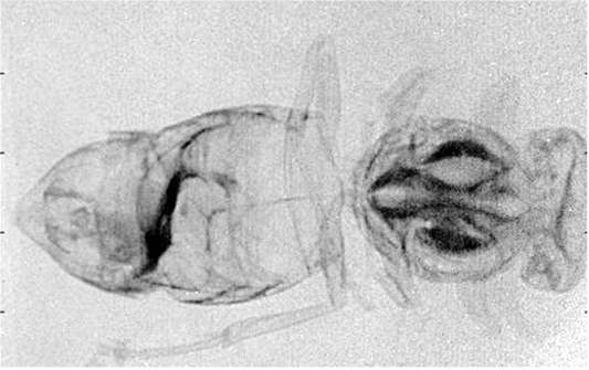
This is a simple propagation-based X-ray phase contrast image of an insect, obtained with a conventional (large spot) x-ray source and a polycapillary optic.
The Center for X-ray Optics specializes in developing x-ray techniques and technology for a variety of applications. A major recent emphasis is on the development of simple techniques to bring practical x-ray phase imaging to clinical and industrial settings. The center was founded in 1991 as a collection of faculty, postdoctoral researchers, graduate and undergraduate students and international collaborators studying the properties and application of x-ray and neutron optics.
Facilities include a 1200 sq ft laboratory housing multiple x-ray beam lines consisting of rotating anode or microfocus x-ray sources with copper, molybdenum and tungsten targets, motion control systems for optic alignment and testing, and high resolution energy sensitive or imaging detectors.
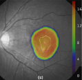
Simulated dose mapping using polycapillary optic for the treatment of macular degeneration.
X rays have been very important in many areas of science and technology since their discovery. The first medical radiograph was made in 1896, just one year after their discovery by Roentgen.
Because most x-ray creation techniques generate x rays which diverge away from their source like light from a light bulb, capturing and collimating, or focusing, x-rays is helpful.
However, because x-rays easily penetrate most objects (even people), they simply pass through normal lenses, hardly bending at all.
Similarly, normal mirrors don't work because the ray simply goes through the mirror without reflecting.
X-ray optics seek to overcome these challenges through the use of reflective optics, polycapillary optics and crystal optics techniques.
Reflective Optics
Reflective Optics
The bending of light by a lens is described by Snell's law, which tells us that when light travels from a material of low index, like air, to a material of higher index, like glass, it bends, or refracts, away from the glass surface.
However, for x rays, the index for glass is actually very slightly less than that of air or vacuum. This means that the ray bends very slightly toward the glass surface. If the x ray hits the surface at a special, very small angle called the critical angle, the ray in the glass will be parallel to the surface.
For incident angles less than the critical angle, there is no way to have a ray in the glass, so the x ray is totally reflected. This principle has been used for many years to make x-ray mirrors. However the critical angle is usually less than one tenth of one degree, and so the mirror has to be very large and very flat to reflect a large x-ray beam.
The same principle has been used since the 1920s to bounce x rays down hollow glass tubes. Of course a single tube can only catch a very small part of the x-ray beam.
Polycapillary Optics
Polycapillary Optics
In the early 1980's polycapillary optics, arrays of thousands of hollow glass tubes, were invented in Russiaby Kumakhov and coworkers. In 1991, the Institute for Roentgen Optics in Moscow, and the Center for X-ray Optics in Albany were jointly founded to purse the study of these and other x-ray optics.
The Center for X-ray Optics now houses a half dozen x-ray beam enclosures, including several microfocus sources, with copper, molybdenum and tungsten tubes, high resolution energy sensitive and imaging detectors, and two high power rotating anode systems used for both imaging and crystallography experiments.
Theoretical development and extensive computer modeling are also a large part of the research program, as well as collaborations with several synchrotron beam lines and neutron sources.
Gaining More Control Over X-rays
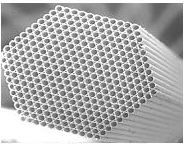
A cross-sectional scanning electron micrograph of a polycapillary fiber with 0.55 mm outer diameter and 50 µm diameter channels.
Polycapillary optics can control x rays over a broad range of angles and energies and have been used as focusing collectors for x-ray astronomy to produce large area collimated beams for wafer analysis, and to provide small focused beams for protein crystallography with low power x-ray sources.
They are also being developed for a number of medical applications, including the removal of Comptonscattering with the resultant improvement in contrast and resolution in mammography, the production of monochromatic parallel beams for high contrast imaging in a clinical setting, and the detection and localization of radioactive tracers in prostate cancer.
Other exciting applications are extensions of measurements normally performed at synchrotrons into laboratory or clinical settings because of the increased efficiency of source utilization.
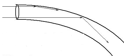
X-rays traveling through a bent capillary tube bounce off the inner tube wall. The lower x-ray, entering closer to the center of curvature, strikes the tube wall at a larger angle.
Polycapillary optics are arrays of hollow glass tubes used to collect, focus, and redirect x-ray and neutron beams. X-rays striking the interior of these hollow channels at grazing incidence are guided along the channel by total external reflection in a process similar to the way fiber optics guide light.
The reflection of x-rays, which are reflected down the length of the capillary, is governed by the critical angle, which is approximately 1.5 mrad or 0.1° at 20 keV and is inversely proportional to photon energy. X-rays are transmitted down in hollow glass tubes with high efficiency so long as the incidence angles are kept smaller than the critical angle.
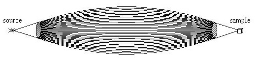
Focusing or collecting effects come from the overlap of the beams from thousands of capillary channels, rather than from the action within a single tube. As for single bore capillaries, x rays can be transmitted down a curved hollow tube as long as the tube is small enough and bent gently enough to keep the angles of incidence less than the critical angle for total reflection, #c.
A sketch of the interior channels of a monolithic polycapillary optic. Monolithic optics can be focusing, as shown, or collimating.
The angle of incidence for the ray near one edge increases with tube diameter. The requirement that the incident angles remain less than the critical angle necessitates the use of tiny tubes. Polycapillary fibers have tube diameters that are much smaller than the fiber diameter, while still maintaining high open area.
Typical channel sizes are between 2 and 12 mm. Thousands of such fibers are strung through lithographically produced metal grids to produce a multifiber lens. Alternatively, a larger diameter polycapillary fiber can be shaped into a one-piece, monolithic, optic.
Because x rays, like all light, are also waves, there can be interference effects. The wavelength of x-rays is on the order of 0.1 nanometer, similar to the spacing between atoms in solids, so there are complex interference effects, called diffraction, when x rays are passed into solids.
This is routinely used to reflect x rays at much high angles than can be achieved by grazing incidence total reflection. However, because it is an interference effect, a crystal "mirror" only works for a single wavelength.
This is a disadvantage if it is desirable to keep most of the intensity from a typical source, which emits a large range of wavelengths, but an advantage for applications in which it is desirable to use a beam of a single wavelength (called monochromatic, since for visible light a single wavelength has a single color).
The Center for X-ray Optics has made use of flat crystal optics since its inception and has been studying curved crystal optics since 2001.
Rohaan Khan, Brenda Adhiambo, Sean Starr-Baier, Danhong Li, Weiyuan Sun, Laila Hassan, C.A. MacDonald, and Jonathan C. Petruccelli, Low dose imaging with simultaneous scatter, attenuation and mesh-based phase contrast,” SPIE Proc. 10948-64, 2019.
Weiyuan Sun, Congxiao He, Carolyn A. MacDonald and Jonathan C. Petruccelli, “Mesh-based and polycapillary optics-based x-ray phase imaging,” SPIE Proc. 10948-176, 2019.
Laila Hassan, Sean Starr-Baier, C. A. MacDonald, Jonathan C. Petruccelli, “Monte Carlo simulations of a novel coherent scatter materials discrimination system,” SPIE Proc. 10187, 1018706-1:9, 2017.
Huagang Yan, Xiangyu Ma, Weiyuan Sun, Stacy Mendez, Stefan Stryker, Sean Starr-Baier, Gianpiero Delliturri, Ravinder Nath, Zhe (Jay) Chen, Kenneth Roberts, Carolyn MacDonald, and Wu Liu, “Monte Carlo dosimetry modeling of focused kV x‐ray radiotherapy of eye diseases with potential nanoparticle dose enhancement,” Medical Physics 45 (10), 8/2018.
W. M. Gibson, C.A. MacDonald, and N. Mail, “Potential for Radioscintography with Polycapillary Optics,” in Ali M. Khounsary , C.A. MacDonald, eds., Advances in Laboratory-Based X-Ray Sources and Optics III, SPIE vol. 4781, pp. 104-111, 2002.
C.A. MacDonald, An Introduction to X-Ray Physics, Optics, and Applications, Princeton University Press, 2017.
C. A. MacDonald, invited review article, “Laboratory x-ray optics,” Annual Review of Materials Research, Volume 47, Novel Functionality Through Metamaterials, pp. 115-134, 2017.
C.A. MacDonald and W.M. Gibson, “An Introduction to X-ray and Neutron Optics,” chapter 19 in M. Bass, ed., Handbook of Optics, Volume III, McGraw-Hill 2000.
J.B. Ullrich and C.A. MacDonald, “Electron Impact Sources,” chapter 31 in M. Bass, ed., Handbook of Optics, Volume III, McGraw-Hill 2000.
C.A. MacDonald and W.M. Gibson, “Applications Requirements Affecting Optics Selection,” chapter 35 in M. Bass, ed., Handbook of Optics, Volume III, McGraw-Hill 2000.
W.M. Gibson and C.A. MacDonald, “Summary of X-ray and Neutron Optics,” chapter 37 in M. Bass, ed., Handbook of Optics, Volume III, McGraw-Hill 2000.
W.M. Gibson, H. Huang, J. Nicolich, P. Klein, and C.A. MacDonald, “Polycapillary Optics For Angular Filtering Of X Rays In Two Dimensions”, in Advances in X-ray Analysis, 45, F-58, Proceedings of the 2001 50th Denver X-ray Conference, 2002.
T. Bievenue, J. Burdett, Z.W. Chen, N. Gao, D.M. Gibson, W.M. Gibson*, H. Huang, and I.Yu. Ponomarev, “New capabilities and applications of compact source-optic combinations,”Ali M. Khounsary and C.A. MacDonald, eds., Advances in Laboratory-Based X-Ray Sources and Optics II, SPIE vol. 4502, pp. 134-47, 2001.
Danhong Li, Noor Mail, C.A. MacDonald, “A comparison of doubly curved crystal and polycapillary optics for monochromatic beam production from a clinical source,” in M. J. Flynn, ed., Physics of Medical Imaging, SPIE vol. 5745, pp.754-763, 2005.
F.R. Sugiro, S. D. Padiyar, C. A. MacDonald, “Characterization of Pre- and Post- Patient X-ray Polycapillary Optics for Mammographic Imaging” in C.A. MacDonald and Ali M. Khounsary, eds., Advances in Laboratory-Based X-Ray Sources and Optics, SPIE vol. 4144, pp. 204-215, 2000.
Cari, C.A. MacDonaldand W.M. Gibson, C. D. Alexander and M.K. Joy, C.H. Russell, Z. W. Chen, “Characterization of a Long Focal Length Polycapillary Optic for High Energy X rays,” in C.A. MacDonald and Ali M. Khounsary, eds., Advances in Laboratory-Based X-Ray Sources and Optics, SPIE vol. 4144, pp.183-192, 2000.
S.D. Padiyar, M.V. Gubarev, Hui Wang, W.M. Gibson, C.A. MacDonald “Characterization of Polycapillary X-Ray Collimating Optics”, in C.A. MacDonald, K.A. Goldberg, J.R. Maldonado, A.J. Marker III, S.P. Vernon, eds., EUV, X-ray, and Neutron Optics and Sources, SPIE vol. 3767, pp. 90-101, 1999.
Lei Wang and C.A. MacDonald, “Measurement of Capillary Optic Performance for Hard X rays,” in X-Ray and Ultraviolet Sensors and Applications, R.B. Hoover and M.B. Williams, eds., SPIE vol. 2519, pp. 218-223, July 1995.
C.A. MacDonald, C.C. Abreu, S. Budkov, H. Chen, X. Fu, W.M. Gibson, Kardiawarman, A. Karnaukhov, V. Kovantsev, I. Ponomarev, B.K. Rath, J.B. Ullrich, M. Vartanian, Q. F. Xiao, "Quantitative Measurements of the Performance of Capillary X-Ray Optics," in Multilayer and Grazing Incidence X-Ray/EUV Optics II, R.B. Hoover and A. Walker, eds., SPIE Proc. vol. 2011, pp. 275-286, 1993.
Simulation and Theory
Hui Wang, Lei Wang, W.M. Gibson, C.A. MacDonald, “Simulation Study oF Polycapillary X-Ray Optics,” in X-Ray Optics, Instruments, and Missions, R.B. Hoover and A.B.C. Walker II, eds. SPIE Vol 3444, pp. 643-651, July 1998.
Lei Wang, B.K. Rath, W.M. Gibson, J.C. Kimball, C.A. MacDonald, "Performance Study of Polycapillary Optic Performance for Hard X rays," Journal of Applied Physics, 80 (7), pp.3628-3638, October 1, 1996.
Lei Wang, B.K. Rath W.M. Gibson, J.C. Kimball, and C.A. MacDonald, "Analysis of capillary optic performance for hard x rays", Hard X-Ray/Gamma-Ray and Neutron Optics, Sensors, and Applications, R.B. Hoover, and F.P. Doty, eds., SPIE Proceedings Vol. 2859, pp. 170-181, 1996.
D. Bittel, and J. Kimball, J. Appl. Phys., 74(2), (1993).
Q.-F. Xiao, I.Yu. Ponomarev, A.I. Kolomitsev and J.C. Kimball, "Numerical Simulations for Capillary-Based X-ray Optics", in X-ray Detector Physics and Applications, (R.B. Hoover, ed.), SPIE Proc. 1736, (1992).
Medical
C.A. MacDonald, J. C. Petruccelli , “Polycapillary optics for medical applications,” Journal of Physics: Conference Series 776 (2016) 012001, doi:10.1088/1742-6596/776/1/012001.
Danhong Li, Noor Mail, C.A. MacDonald, “A comparison of doubly curved crystal and polycapillary optics for monochromatic beam production from a clinical source,” in M. J. Flynn, ed., Physics of Medical Imaging, SPIE vol. 5745, pp.754-763, 2005.
C.A. MacDonald, Noor Mail, W.M. Gibson, SM Jorgensen, E L Ritman, “Micro gamma camera optics with high sensitivity and resolution”, in M. J. Flynn, ed., Physics of Medical Imaging, SPIE vol. 5745, pp.1-6, 2005.
F.R. Sugiro, Danhong Li, C.A.MacDonald, “Beam Collimation with Polycapillary X-ray Optics for High Contrast High Resolution Monochromatic Imaging,”, Med. Phys., 31, p. 3288, 2004.
Jorgensen SM, Chmelik MS, Eaker DR, MacDonald CA, Ritman E L A “Polycapillary X-ray Optics-based Integrated micro-SPECT/CT Scanner,” in Developments in X-ray Tomography IV, Ulrich Bonse, ed., SPIE vol. 5535, pp 36-42, 2004.
D. Li, F.R. Sugiro, C.A. MacDonald, “Source-optic optimization for compact monochromatic imaging”, in X-ray Sources and Optics, C.A. MacDonald, A.T. Macrander, T. Ishikawa,C. Morawe, J.L. Wood, eds., SPIE vol. 5537, pp. 105-114, 2004.
C.A. MacDonald, N. Mail, F. Sugiro, D. Li, “Monochromatic Applications of Polycapillary Optics”, in Laser-Generated and Other Laboratory X-Ray and EUV Sources, Optics, and Applications, George A. Kyrala, Jean-Claude J. Gauthier, Carolyn A. MacDonald, Ali M. Khounsary, eds., SPIE vol. 5196, 2003.
W. M. Gibson, C.A. MacDonald, and N. Mail, “Potential for Radioscintography with Polycapillary Optics,” in Ali M. Khounsary , C.A. MacDonald, eds., Advances in Laboratory-Based X-Ray Sources and Optics III, SPIE vol. 4781, pp. 104-111, 2002.
C.A. MacDonald, W.M. Gibson, and W. Peppler, “X-Ray Optics for Better Diagnostic Imaging,” Technology In Cancer Research And Treatment, 1, (2), April 2002, pp 111-118.
C. A. MacDonald, W. M. Gibson, F. R. Sugiro, “High Contrast Imaging With Polycapillary Optics,” Advances in X-ray Analysis, 45, Proceedings of the 50th Denver X-ray Conference, in press.
C.A.MacDonald,F.R. Sugiro, and W.M. Gibson, “Improved Radiography with Polycapillary X-Ray Optics,” Ali M. Khounsary and C.A.MacDonald, eds., Advances in Laboratory-Based X-Ray Sources and Optics II, SPIE vol. 4502, pp. 10-18, 2001.
F.R. Sugiro and C.A. MacDonald, “Monochromatic Imaging with a Conventional Source Using Polycapillary X-ray Optics,” in L.E. Antonuk, M.J. Yaffe, eds., Medical Imaging 2001:Physics of Medical Imaging, SPIE vol. 4320, pp. 427-434, 2001.
Cari, Suparmi, W. M. Gibson, C. A. MacDonald, “Contrast Enhancement Measurements Using Polycapillary X-Ray Optics at 20-40 keV,” in L.E. Antonuk, M.J. Yaffe, eds., Medical Imaging 2001:Physics of Medical Imaging, SPIE vol. 4320, pp.163-170, 2001.
F.R. Sugiro, S. D. Padiyar, C. A. MacDonald, “Characterization of Pre- and Post- Patient X-ray Polycapillary Optics for Mammographic Imaging” in C.A. MacDonald and Ali M. Khounsary, eds., Advances in Laboratory-Based X-Ray Sources and Optics, SPIE vol. 4144, pp. 204-215, 2000.
Suparmi, Cari, W.M. Gibson and C.A. MacDonald, “Development of Polycapillary X-Ray Optics for Scatter Rejection,” inC.A. MacDonald and Ali M. Khounsary, eds., Advances in Laboratory-Based X-Ray Sources and Optics, SPIE vol. 4144, pp.216-227, 2000.
S.M. Jorgensen, D.A. Reyes, C.A. MacDonald, E.L. Ritman, “Micro-CT Scanner With A Focusing Polycapillary X-Ray Optic,”in U. Bonse, ed., Developments in X-Ray Tomography II, SPIE vol. 3772, 1999, pp. 158-166.
Lei Wang, W.M. Gibson, C.A. MacDonald, “Potential of Polycapillary X-Ray Optics in Medical Imaging Applications,”in C.A. MacDonald, K.A. Goldberg, J.R. Maldonado, A.J. Marker III, S.P. Vernon, eds., EUV, X-ray, and Neutron Optics and Sources, SPIE vol. 3767, pp.102-112, 1999.
C.C. Abreu and C.A. MacDonald, “Beam Collimation, Focusing, Filtering and Imaging with Polycapillary X-ray and Neutron Optics,” review article, Physica Medica, vol. XIII, N.3, 1997, pp. 79-89.
D.G. Kruger, C.C. Abreu, E.G. Hendee, A. Kocharian, W.W. Peppler, C.A.Mistretta, C.A.MacDonald, “Imaging Characteristics of X-Ray Capillary Optics in Mammography,” Medical Physics 23 (2), pp. 187-196, February 1996.
C.C. Abreu, D.G. Kruger, C.A. MacDonald, C.A. Mistretta, W.W. Peppler, Q.F. Xiao, "Measurements of Capillary X-Ray Optics with Potential for Use in Mammographic Imaging," Medical Physics 22 (11), Pt. 1, pp. 1793-1801, November 1995.
C.A. MacDonald and W.M. Gibson, “Medical Applications of Polycapillary X-Ray Optics,” in X-Ray and Ultraviolet Sensors and Applications, R.B. Hoover and M.B. Williams, eds.,SPIE vol. 2519, pp. 186-196, July 1995.
P. Tompkins, C.C. Abreu, F. Carrol, Q. Xiao, C.A.MacDonald, "Use of Capillary Optics as a Beam Intensifier for a ComptonX-Ray Source," Medical Physics, 21, no. 11, pp. 1777-1784, 1994.
W.M. Gibson, C.A. MacDonald, and M.S. Kumakhov, "The Kumakhov Lens; A New X-Ray and Neutron Optics with Potential for Medical Applications," in Technology Requirements for Biomedical Imaging, S. K. Mun, ed., I.E.E.E. Press #2580, 1991, pp.164-169.
Synchrotron Focusing/Radiation Damage
B.K. Rath, W.M. Gibson, Lei Wang, B.E. Homan and C.A. MacDonald, “Measurement and Analysis of Radiation Effects in Polycapillary X-ray Optics,” Journal of Applied Physics, 83, no.12, pp. 7424-7435, June 15 1998.
B. K. Rath, F. B. Hagelberg, B. E. Homan and C. A. MacDonald, “Synchrotron White Beam Thermal Loading on Polycapillary X-ray Optics,” Nuclear Instruments and Methods A, vol. 401, nos. 2,3, pp. 421--428, 1997.
F.A. Hofmann, C.A. Freinberg-Trufas, S.M. Owens, S.D. Padiyar, C.A. MacDonald, “Focusing of Synchrotron Radiation with Polycapillary Optics,” Beam Interactions with Materials and Atoms: Nuclear Instruments and Methods B, vol. 133, 1997, pp. 145-150.
B.K. Rath, D.C. Aloisi, D.H. Bilderback, N. Gao, W.M. Gibson, F.A. Hofmann, B.E. Homan, C.J. Jezewski, I.L. Klotzko, J.M. Mitchell, S.M. Owens, J.B. Ullrich, Lei Wang, G.M. Wells, Q.F. Xiao, and C.A. MacDonald, “Effects of intense x-ray radiation on polycapillary fiber performance,” in X-Ray and Ultraviolet Sensors and Applications, R.B. Hoover and M.B. Williams, eds., SPIE vol. 2519, pp. 207-217, July 1995.
Q.-F. Xiao, I.Yu. Ponomarev, A.I. Kolomitsev, D.M. Gibson, F.A. Dilmanian, and E. Nachaliel, "Guidance of Hard X-rays using Glass Polycapillary Fiber", Proc. of 8th Annual Conf. on Synchrotron Radiation Instrumentation, NIST, Gaithersburg, MD, October, 1993, Nucl. Inst. and Methods in Physics Res. (1994)
J.B. Ullrich, W.M. Gibson, M.V. Gubarev, C.A.MacDonald, "Potential for Concentration of Synchrotron Beams with Capillary Optics," Nuclear Instruments and Methods in Physics Research A 347, pp. 401-406, 1994.
V.A. Arkadiev, R. Fayazov, M.A. Kumakhov and W.M. Gibson, "Concentration of Synchrotron Radiation with Capillary Arrays", in Optics for High Brightness Synchrotron Radiation Beamlines, SPIE Proc. (1992).
Diffraction
N. Mail, W. M. Gibson, and, C.A. MacDonald, “Molybdenum Microfocus Source Coupling to Polycapillary Optics for Powder Diffraction,” in Ali M. Khounsary , C.A. MacDonald, eds., Advances in Laboratory-Based X-Ray Sources and Optics III, SPIE vol. 4781, pp. 87-95, 2002.
H. Huang, C. A. MacDonald, W. M. Gibson, J.R. Ruble, J. X. Ho, J. Chik, A. Parsegian, and I. Ponomarev, “Focusing Polycapillary Optics For Diffraction,” to be published in Advances in X-ray Analysis, 45, Proceedings of the 50thDenver X-ray Conference, 2001.
H. Huang, C. A. MacDonald, W. M. Gibson, J. Chik, A. Parsegian, and I. Ponomarev, “Collimating And Focusing Polycapillary Optics For Powder Diffraction”, Ali M. Khounsary and C.A. MacDonald, eds., Advances in Laboratory-Based X-Ray Sources and Optics II, SPIE vol. 4502, pp.30-37, 2001.
H. Huang, C.A.MacDonald, W.M. Gibson, D.C. Carter, J.X. Ho, J.R. Ruble, I. Ponomarev, “Low Power Protein Crystallography using Polycapillary Optics” in C.A. MacDonald and Ali M. Khounsary, eds., Advances in Laboratory-Based X-Ray Sources and Optics, SPIE vol. 4144, pp.100-109, 2000.
S.D. Padiyar, H. Wang, W.M. Gibson, C.A. MacDonald, M.V. Gubarev, “Beam Collimation Using Polycapillary X-Ray Optics For Large Area Diffraction Applications,” in Advances in X-ray Analysis, 43, Proceedings of the 48th Denver X-ray Conference, 1999.
F.A. Hofmann, W.M. Gibson, C.A. MacDonald, D.A. Carter, J.X. Ho, J.R. Ruble, “Low Power Polycapillary Based System For X-Ray Protein Crystallography,” in Advances in X-ray Analysis, 43, Proceedings of the 48th Denver X-ray Conference, 1999.
W.M. Gibson, C.A. MacDonald, J.B. Ulllrich, "Diffraction Geometry Optimization with Polycapillary X-Ray Optics", in C.A. MacDonald, K.A. Goldberg, J.R. Maldonado, A.J. Marker III, S.P. Vernon, eds., EUV, X-ray, and Neutron Optics and Sources, SPIE vol. 3767, pp. 199-208, 1999.
C.A. MacDonald, S.M. Owens, and W.M. Gibson, "Polycapillary X-Ray Optics for Microdiffraction," Journal of Applied Crystallography, 32, pp160-7, 1999.
F.A. Hoffman, N. Gao, S.M. Owens, W.M. Gibson, C.A. MacDonald, and S.M. Lee, “Polycapillary Optics For In-Situ Process Diagnotstics,” P.A. Rosenthal, W.M. Duncan, J.A. Woollam, eds., In Situ Process Diagnostics and Intelligent Materials Processing, Materials Research Society Proceedings, vol. 502, pp 133-138, 1998.
F. A. Hofmann, W.M. Gibson, S.M. Lee, C.A. MacDonald, “Polycapillary X-Ray Optics For Thin Film Strain and Texture Analysis,” in Thin Film Stresses and Mechanical Properties VII, R.C. Cammarata, M.A. Nastasi, E.P. Busso, W.C. Oliver, eds., Materials Research Society Proceedings, vol. 505, 1998.
S.M. Owens, F.A. Hoffman, C.A. MacDonald and W.M. Gibson, " Microdiffraction using Collimating and Convergent Beam Polycapillary Optics", , Advances in X-ray Analysis, Volume 41, Proceedings of the 46th Annual Denver X-ray Conference Proceedings, Steamboat Springs, Colorado, August 4-8, 1997.
Kardiawarman, B.R. York, X.W. Qian, Q.F. Xiao, C.A. MacDonald, and W.M. Gibson,“Application of a Multifiber Collimating Lens to Thin Film Structure Analysis,” X-Ray and Ultraviolet Sensors and Applications, in R.B. Hoover and M.B. Williams, eds., SPIE vol. 2519, pp.197-206, July 1995.
J.B. Ullrich, I.Yu. Ponomarev, M.V. Gubarev, N. Gao, Q.-F. Xiao and W.M. Gibson, "Development of Monolithic Capillary Optics for X-ray Diffraction Applications", SPIE Ann. Mtng. San Diego, CA, July, 1994.
Kardiawarman, V. Kovantsev, S. Budkov, Q.F. Xiao, W.M. Gibson, C.A. MacDonald, P. Persans, L. Lurio, ‘Characterization of a Multi-Fiber Polycapillary-based X-Ray Collimating Lens,” in X-Ray and UV Detectors, SPIE vol. 2278, pp. 238-247, 1994.
Microfluorescence
Y. Yan and W. Gibson, Polycapillary Optics for Analytical Analysis, Denver 2001
F.A. Hoffman, N. Gao, S.M. Owens, W.M. Gibson, C.A. MacDonald, and S.M. Lee, “Polycapillary Optics For In-Situ Process Diagnotstics,” P.A. Rosenthal, W.M. Duncan, J.A. Woollam, eds., In Situ Process Diagnostics and Intelligent Materials Processing, Materials Research Society Proceedings, vol. 502, pp 133-138, 1998.
D.A. Wollman, C. Jezewiski, G.C. Hilton, Q.F. Xiao, K.D. Irwin, L.L. Dulcie, and J.M. Martinis, Proc. Microscopy and Microanalysis, 3, 1075 (1997)
N. Gao, I.Yu. Ponomarev, Q.F. Xiao, and W.M. Gibson, Appl. Phys. Lett. 71, 3441-3 (1997)
N. Gao, I.Yu. Ponomarev, Q.F. Xiao, W.M. Gibson, and D.A. Carpenter, Appl. Phys. Lett. 69, 1529 (1996)
Astronomy
C.H. Russell, M. Gubarev, J. Kolodziejczak, M. Joy, C.A. MacDonald, and W.M. Gibson, “Polycapillary X-ray Optics for X-ray Astronomy,” in Advances in X-ray Analysis, 43, Proceedings of the 48th Denver X-ray Conference, 1999.
M.V. Gubarev, C. Bankston, M. Joy, J. Kolodziejczak, C.A. MacDonald, C.H. Russell, W.M. Gibson, “Experimental investigation of the x-ray capillary system for hard x-ray astronomy,” in X-Ray Optics, Instruments, and Missions, R.B. Hoover and A.B.C. Walker II, eds. SPIE Vol 3444, pp. 467-478, July 1998.
C.H. Russell, W. M. Gibson, M. V. Gubarev, F. A. Hofmann, M. K. Joy, C. A. MacDonald, Lei Wang, Qi-Fan Xiao, R. Youngman, “Application of Polycapillary Optics for Hard X-Ray Astronomy” (Invited Paper), in Grazing Incidence and Multilayer X-ray Optical Systems, R.B. Hoover and A.B.C. Walker II, eds., SPIE vol. 3113, pp. 369-377, 1997.
Lithography
M. Vartanian, D.M. Gibson, R. Frankel and J. Drumheller, " Development of a Polycapillary Collimator for Point Source X-ray Lithography", SPIE Proc., 1924, 335-345 (1993).
M. Vartanian, R. Youngman, D. Gibson, J. Drumheller, and R.D. Frankel, "Polycapillary Collimator for Point Source Proximity X-ray Lithography", J. Vac. Sci. Technol. B, 11, (1993).
Doubly Curved Crystal
General
N. Mail, Z. W. Chen, W. M. Gibson, C. A. MacDonald, “Application of Doubly-Curved Crystal Optics,” in X-ray Sources and Optics, C.A. MacDonald, A.T. Macrander, T. Ishikawa,C. Morawe, J.L. Wood, eds., SPIE vol. 5537, pp. 94-104, 2004.
Fluorescence
Z. W. Chen, W. M. Gibson, N. Mail, C. A. MacDonald, “Total reflection x-ray fluorescence with low power sources coupled to DCC optics,” accepted Spectrachemica. Acta.
Medical
Danhong Li, Noor Mail, C.A. MacDonald, “A comparison of doubly curved crystal and polycapillary optics for monochromatic beam production from a clinical source,” in M. J. Flynn, ed., Physics of Medical Imaging, SPIE vol. 5745, pp.754-763, 2005
Neutron Optics
Neutron Optics
Neutron Optics
W.M. Gibson, H.H. Chen-Mayer, D.F.R. Mildner, H.J. Prask, A.J. Schultz, R. Youngmana, T. Gnäupel-Herold, M.E. Miller, and R. Vitt, Polycapillary Optics Based Neutron Focusing For Small Sample Neutron Crystallography, DX 2001
H. Chen, D.F.R. Mildner, R.G. Downing, R.E. Benenson, Q.-F. Xiao and V.A. Sharov, Nucl. Instrum. and Methods, B, (1994).
Q.F. Xiao, H. Chen, D.F.R. Mildner, R.G. Downing and R.E. Benenson, Rev. Sci. Instrum. 64, 3252-3257 (1993).
H. Chen, R.G. Downing, D.F.R. Mildner, W.M. Gibson, M.A. Kumakhov, I.Yu. Ponomarev, and M.V. Gubarev, "Guiding and Focusing Neutron Beams using Capillary Optics", Nature, 357, 391-393.
Detectors
Detectors
Detectors
C. Freinberg-Trufas, W.M. Gibson, S.M. Owens, C. H. Russell, C.A. MacDonald, F. P. Doty, “Measurements of a Prototype CdZnTe Array for Direct Digital Mammography,” in Hard X-ray and Gamma-ray Detector Physics Optics and Applications, R.B. Hoover, F
D.A. Wollman, C. Jezewiski, G.C. Hilton, Q.F. Xiao, K.D. Irwin, L.L. Dulcie, and J.M. Martinis, Proc. Microscopy and Microanalysis, 3, 1075 (1997)
R. Wentink, J. Carbone, D. Aloisi, W.M. Gibson, C.A. MacDonald, Q.E. Hanley, R.E. Fields, and M.B. Denton, “Charge Injection Detectors for X-Ray Imaging,” in Advances in Multilayer and Grazing Incidence X-ray/EUV/FUV Optics, R.B. Hoover and A.B.C. Walker, Jr., eds., Proceedings of the SPIE vol. 2279, 1994, pp. 380-387.
W.M. Gibson, G.L. Miller, and P.F. Donovan, in “Alpha, Beta, and Gamma Spectroscopy,” 2nd ed., K. Siegbahn, ed. (North Holland Publishing Co, Amsterdam, 1964)
G.L. Miller, W.M. Gibson, and P.F. Donovan, Ann. Rev. Nucl. Sci., 33, 380 (1962)
Rohaan Khan, Brenda Adhiambo, Sean Starr-Baier, Danhong Li, Weiyuan Sun, Laila Hassan, C.A. MacDonald, and Jonathan C. Petruccelli, Low dose imaging with simultaneous scatter, attenuation and mesh-based phase contrast,” SPIE Proc. 10948-64, 2019.
Weiyuan Sun, Congxiao He, Carolyn A. MacDonald and Jonathan C. Petruccelli, “Mesh-based and polycapillary optics-based x-ray phase imaging,” SPIE Proc. 10948-176, 2019.
Huagang Yan, Xiangyu Ma, Weiyuan Sun, Stacy Mendez, Stefan Stryker, Sean Starr-Baier, Gianpiero Delliturri, Ravinder Nath, Zhe (Jay) Chen, Kenneth Roberts, Carolyn MacDonald, and Wu Liu, “Monte Carlo dosimetry modeling of focused kV x‐ray radiotherapy of eye diseases with potential nanoparticle dose enhancement,” Medical Physics 45 (10), 8/2018.
2017
2017
C.A. MacDonald, An Introduction to X-Ray Physics, Optics, and Applications, Princeton University Press, 2017.
C. A. MacDonald, invited review article, “Laboratory x-ray optics,” Annual Review of Materials Research, Volume 47, Novel Functionality Through Metamaterials, pp. 115-134, 2017.
Laila Hassan, Sean Starr-Baier, C. A. MacDonald, Jonathan C. Petruccelli, “Monte Carlo simulations of a novel coherent scatter materials discrimination system,” SPIE Proc. 10187, 1018706-1:9, 2017.
Katie Kern, Laila Hassan, Lubna Peerzada, Mahboob Ur-Rehman, Carolyn A. MacDonald, "Measurements and simulations of scatter imaging as a simultaneous adjunct for screening mammography", in Medical Imaging 2015: Physics of Medical Imaging, Christoph Hoeschen; Despina Kontos, Editors, Proceedings of SPIE Vol. 9412, 941241, 2015.
Jonathan C. Petruccelli, Sajid Bashir, Sajjad Tahir, Bushra Kanwal, Carolyn A. MacDonald, "Practicable phase contrast techniques with large spot sources", in Medical Imaging 2015: Physics of Medical Imaging, Christoph Hoeschen; Despina Kontos, Editors, Proceedings of SPIE Vol. 9412, 941254, 2015.
Ali M. Khounsary, Carolyn A. MacDonald, eds., Advances in Laboratory-based X-Ray Sources, Optics, and Applications IV, SPIE proc. 9590, 2015.
Jonathan C. Petruccelli, Sajjad Tahir, Sajid Bashir, Adam Pan, Lei Tian, Ling Xu, Carolyn A. MacDonald, George Barbastathis, Computational x-ray phase imaging, in Proc. SPIE 9209, Advances in Computational Methods for X-Ray Optics III, 92090P, 2014.
Sajid Bashir, Jonathan C. Petruccelli, Carolyn MacDonald, Phase imaging using polycapillary optics Proc. SPIE 9207, Advances in X-Ray/EUV Optics and Components IX, 92070X, 2014.
Laila Hassan, Lubna Peerzada, Katie Kern, C. A. MacDonald, “Coherent scatter imaging simulation for screening mammography,” in Penetrating Radiation Systems and Applications XIII, Proceedings of SPIE Volume: 8509, 2012.
2010
M. Bass, C. DeCusatis, J Enoch, V. Lakshminarayanan, G. Li, C. MacDonald, V.N. Mahajan, E. VanStryland, eds., Handbook of Optics Third Ed., Volumes I-V, McGraw-Hill 2010.
Ayhan Bingolbali and C. A. MacDonald, “Quality assessment system for curved crystal X-ray optics," Nuclear Instruments and Methods in Physics Research Section B: Beam Interactions with Materials and Atoms, Volume 267, Issue 5, March 2009, Pages 832-841.
D.G. Kruger, C.C. Abreu, E.G. Hendee, A. Kocharian, W.W. Peppler, C.A. Mistretta, C.A. MacDonald, “Imaging Characteristics of X-Ray Capillary Optics in Mammography,” Medical Physics 23 (2), pp. 187-196, February 1996.
C.A. MacDonald, "Applications and Measurements of Polycapillary X-Ray Optics," Invited Paper, Journal of X-Ray Science and Technology, 6, pp. 32-47, 1996, PMID 21307511
B. Rath, R. Youngman, C.A. MacDonald, "An Automated Test System for Measuring Polycapillary X-Ray Optics," Review of Scientific Instrumentation, 65, pp.3393-3398, Nov. 1994.
J.B. Ullrich, W.M. Gibson, M.V. Gubarev, C.A. MacDonald, "Potential for Concentration of Synchrotron Beams with Capillary Optics," Nuclear Instruments and Methods in Physics Research A 347, pp. 401-406, 1994.
C.A. MacDonald, A.M. Malvezzi, and F. Spaepen, "Picosecond Time-Resolved Measurements of Crystallization in Noble Metals," Journal of Applied Physics, 65, no. 1, 1988, pp. 129-136.