MELENDEZ RESEARCH GROUP

Melendez Research Group

Reactive Species Control Disease

Mitochondrea
Mitochondria are a primary source of reactive oxygen species that are involved in many disease pathologies

Our studies revolve around a key central paradigm, that oxidant signaling is precise, compartmentalized and amenable to targeted‐antioxidant based therapies. Reactive oxygen and nitro gen species (ROS/RNS), in addition to their ability to damage biomolecules, have also emerged as key mediators in regulation of signaling networks by modulating phosphatase activity, kinase cascades and transcription factor binding. Thus, ROS/RNS serve a dual role, at low concentrations they are secondary signaling molecules that regulate the expression of a wide array of signaling networks, and at high concentrations damage lipids, protein and DNA. The principle mediator of ROS‐dependent signaling is the 2e‐ reduction product of oxygen, H2O2, which is produced in response to numerous physiologic stimuli.

Our work is focused on defining how ROS/RNS drive cellular signaling events that control cellular senescence, metastatic disease, matrix destruction and the virulence of infectious bacteria. We have developed many cutting-edge tools to monitor oxidant production from cells in real time. Our studies indicate that that augmented free radical production initiate or drive age-associated disease. CNSE is unique and provides the scientific infrastructure for the development of innovative therapeutic and diagnostic technologies to limit degenerative disease.


Projects

Scope

Evaluate the role of oxidants in the control of cancer, aging and infectious disease.

Goals

Provide R&D for the development of targeted antioxidant based therapies for the treatment of metastatic cancer, aging and infection disease.

Project 1: Redox control of senescence and disease
About
Senescence Associated production of mitochondrial H2O2
The senescence associated (SA) production of mitochondrial H2O2 governs the amplitude of SASP through phosphatase inhibition enhancing mTOR-signaling, SASP post-transcriptional processing and promoting renal fibrosis.

Fifteen percent of American adults (>30 million) suffer from some degree of chronic kidney disease (CKD). Medicare costs for patients aged 65 years or older with CKD were about $45 billion in 2012. Globally from 8-16% of the population worldwide is affected by CKD and in 2014 kidney disease was the 9th ranked cause of death nationally. Strategies to reduce burden and medical costs related to renal disease are critically needed. Senescence cells have recently emerged as contributors to age-related renal pathology. While cellular senescence has evolved as a protective mechanism to arrest cells exposed to oncogenic insult, chronic senescence activation promotes loss of renal function. The harmful effects of senescence are attributed to high secretory activity, commonly referred to as the Senescence Associated Secretory Phenotype (SASP).

Strategies which limit the amplitude and duration of SASP will serve to delay age related renal decline. SASP activation is reliant on production of interleukin-1 alpha (IL-1α) and we have shown that H2O2, likely of mitochondrial origin, regulates IL-1α transcription and processing in this process. H2O2 signals, in part, through oxidative inactivation of specific protein tyrosine phosphatases (PTPs) that coordinate a broad array of signaling networks. Conversely, IL-1α mRNA stabilization and translation in SASP is also regulated by mTOR. Hence, we have been defining the interplay between H2O2 and PTPs that govern mTOR signaling and how this contributes to gene silencing and post-transcriptional processing of the SASP. Fibrotic insult is a precursor to loss of renal function and we are developing targeted strategies which restrict mitochondrial H2O2, mTOR or SASP and can limit renal fibrosis.

Related Publications
  1. McCarthy, D. a. et al. Redox-control of the alarmin, Interleukin-1α. Redox Biol. 1, 218–225 (2013).
  2. Kar, S., Subbaram, S., Carrico, P. M. & Melendez, J. A. Redox-control of matrix metalloproteinase-1: a critical link between free radicals, matrix remodeling and degenerative disease. Respir. Physiol. Neurobiol. 174, 299–306 (2010).
  3. McCarthy, D. A. et al. Featured Article: Nanoenhanced matrix metalloproteinase-responsive delivery vehicles for disease resolution and imaging. Exp. Biol. Med. 241, (2016).
  4. Bartling, T. R. et al. Redox-sensitive gene-regulatory events controlling aberrant matrix metalloproteinase-1 expression. Free Radic. Biol. Med. 74, (2014).
  5. Sosa Peña, M. del P., Lopez-Soler, R. & Melendez, J. A. Senescence in chronic allograft nephropathy. Am. J. Physiol. Physiol. 315, F880–F889 (2018).
  6. Chandrasekaran, A., Idelchik, M. D. P. S. & Melendez, J. A. Redox control of senescence and age-related disease. Redox Biol. 11, 91–102 (2017).
  7. Dasgupta, J. et al. Reactive oxygen species control senescence-associated matrix metalloproteinase-1 through c-Jun-N-terminal kinase. J. Cell. Physiol. 225, 52–62 (2010).
  8. Nelson, K. K. & Melendez, J. A. Mitochondrial redox control of matrix metalloproteinases. Free Radic. Biol. Med. 37, 768–84 (2004).
  9. Ranganathan, A. C. et al. Manganese Superoxide Dismutase Signals Matrix Metalloproteinase Expression via H2O2-dependent ERK1/2 Activation. J. Biol. Chem. 276, (2001).
  10. Dasgupta, J., Kar, S., Remmen, H. Van & Melendez, J. A. Age-dependent increases in interstitial collagenase and MAP Kinase levels are exacerbated by superoxide dismutase deficiencies. Exp. Gerontol. 44, 503–510 (2009).
  11. Kar, S., Subbaram, S., Carrico, P. M. & Melendez, J. A. Redox-control of matrix metalloproteinase-1: A critical link between free radicals, matrix remodeling and degenerative disease. Respir.Physiol Neurobiol. (2010).
Project 2: Protease-sensing nanoparticles
About
Components of Nanoenhanced MMP-Responsive Delivery Vehicle
Components of Nanoenhanced MMP-Responsive Delivery Vehicle (NMRDV). The image depicts the basic components of NMRDVs. In this study, the matrix components were type I collagen particles, the cargo was either antioxidants (Trolox, Didox, pomegranate extract, apple peel extract, catalase) or the MMPI Ilomostat, and the enhancing FNPs were either GFP-NP, FeOx-NPs, or TRITC-NP

The wide array of proteases, including matrix metalloproteinases (MMPs), produced in response to many pathogenic insults, confers a unique proteolytic signature which is often disease specific and provides a potential therapeutic target for drug delivery. We have been testing the use of Collagen-based Nanoenhanced MMP-Responsive Delivery Vehicles (NMRDVs) that display MMP specific degradation in diverse in vitro models of proteolysis. We demonstrate that collagen particles comprised of protease substrates (primarily collagen) can be made of uniform size and loaded efficiently with assorted cargo including fluorescently-labeled mesoporous silica, magnetic nanoparticles, proteins and antioxidants. We also demonstrate that pathologic concentrations of proteases produced in situ or in vitro display protease specific cargo release. Additionally, we show that the collagen-based particles display bright fluorescence when loaded with a fluorophore, and have the potential to be used as vehicles for targeted delivery of drugs or imaging agents to regions of high proteolytic activity.

Related Publications
  1. McCarthy, D. A. et al. Featured Article: Nanoenhanced matrix metalloproteinase-responsive delivery vehicles for disease resolution and imaging. Exp. Biol. Med. 241, (2016).
  2. Flaherty, N. L. et al. Comparative analysis of redox and inflammatory properties of pristine nanomaterials and commonly used semiconductor manufacturing nano-abrasives. Toxicol. Lett. 239, 205–215 (2015).
Project 3: Selenium, senescence and renal cancer
About
Selenium, senescence and renal cancer

Maintenance of the GSH redox cycle is reliant on the activities of selenocysteine-containing GSH metabolizing enzymes. Selenocysteine is the 21st amino acid and does not contain a dedicated codon. Selenocysteine incorporation during translation requires UGA-stop-codon recoding, which uses specifically modified tRNA for accurate decoding. Dynamic changes in tRNA modification are an epitranscriptomic signal because they regulate gene expression post-transcriptionally. The Begley lab has shown that the stress-induced translation of many selenocysteine containing ROS detoxifying enzymes is dependent on the Alkbh8 tRNA methyltransferase. Alkbh8 enzymatically methylates the uridine wobble base on tRNASelenocysteine to promote UGA-stop codon decoding. Surprisingly the Alkbh8-deficient (Alkbh8-/-) mice reproduce, thrive normally and live past 15 months, suggesting they adapt to the selenoprotein deficiency, high ROS and increased DNA damage levels. In collaboration with the Begley lab we have been investigating a potential adaption mechanism, we have used molecular, biochemical and genomic approaches to demonstrate that Alkbh8-/- mouse embryonic fibroblasts (MEFs) and some organs display markers of senescence and a senescence gene signature. Using theAlkbh8-/- mice we are testing the hypothesis that senescence occurs in vivo as a result of defective epitranscriptomic signals that controls selenocysteine utilization and prevents tumor emergence.

Related Publications
  1. Endres, L. et al. Alkbh8 Regulates Selenocysteine-Protein Expression to Protect against Reactive Oxygen Species Damage. PLoS One 10, e0131335 (2015).

People

Professor Andre Melendez- Principal Investigator

Andres “Andre” Melendez earned his B. Sc. in Marine Science & Biology at the University of Tampa which was followed by an M. Sc. in Biomedical Sciences at the College of Veterinary Medicine of Virginia Polytechnic Institute and State University where he was indoctrinated into the field of free radical biology. He then transitioned to the State University of New York at Albany where he received a Ph. D. in molecular biology and evaluated how reactive oxygen species contribute to inflammatory disease processes. He completed Post-doctoral training at Georgetown University and Albany Medical College where he studied inflammatory processes that accompany oxygen toxicity and post-partum uterine involution, respectively. In 1997 he was one of the first recipients of an NCI Mentored Career Development Award to Promote Diversity. At Albany Medical College he developed a program directed at understanding the free radical signals that control metastatic disease progression, aging and infectious disease processes and funded by both private and federal funding agencies. In 2011, he joined the College of Nanoscale Science & Engineering at the University of Albany, State University of New York as empire innovation professor & associate head of the nanobioscience constellation. He continues his work on free radical signaling and in the development of next generation nanoparticle-based therapeutic vehicles for the diagnosis and treatment of degenerative disease. He is an associate editor for the bionanoscience section of Experimental Biology & Medicine and serves on the editorial board of Free Radical Biology & Medicine and The FASEB Journal. Dr. Melendez has received numerous awards and has served on many review and advisory boards for the government, academic institutions, scientific societies and companies.


May Y. Lee

PhD Student

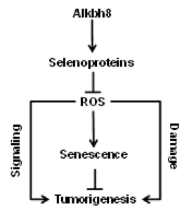
My undergraduate at UAlbany research focused on the effect of a novel tRNA modification by Alkbh8, tRNA methyltransferase enzyme, in aging. As numbers of elderly population are growing dramatically, aging and age-related diseases, such as Alzheimer’s disease, cardiovascular, diabetes and even cancer, will be major public health concerns. Research in aging field will provide major contributions to reduce the burden of age associated illness, and enhance quality of life by maintaining health among older adults population. Thus, I became interested in understanding the intricate web of interdependent genetic, biochemical and physiological factors of aging and age-related diseases. After completing my undergraduate program, I decided to strengthen my research skills by pursuing Master of Science degree, which allow me to continue my research from undergraduate.


Undergraduate (Current)

Habben Desta -- SUNY POLY

Aezat Ullah – Ualbany


Former Graduate Students (Graduated)

Doctoral Thesis Committee

Ana Rodriquez: 1999, “Targeted expression of mitochondrial catalase restricts fibrosarcoma growth in vitro and in vivo.” University of Extremadura, Spain
Current Position: Staff Scientist at King's College London

Aparna Ranganathan: 2002, “ Redox control of Interleukin-1 alpha expression” Albany Medical College
Current Position: Assistant Professor, Basic Medical Sciences, University of Arizona College of Medicine, Phoenix, Arizona

Jenny S. Shum: 2002, “Mechanism behind serotonin dependent control of uterine smooth muscle cell collagenase expression” Albany Medical College
Current Position: Associate, Jones Day, Patent Attorney.

Kristin Nelson: 2004, “Transcriptional control mechanisms regulating hydrogen peroxide-dependent matrix metalloproteinase- 1 expression” Albany Medical College.
Current Position: Medical Writer, University of Louisville, Louisville, Kentucky.

Kip Connor: 2005, “Mitochondrial Hydrogen Peroxide control tumor angiogenesis through PTEN oxidation” Albany Medical College
Current Position: Assistant Professor, Harvard Medical School.

Sita Subbaram: 2008, “Chromatin remodeling events controlling redox-dependent MMP-1 transcription”, Albany Medical College
Current Position: Instructor, Albany Medical College

Jaya Dasgupta: 2008, “Redox-control of senescence associated MMP-1 expression”, Albany Medical College.
Current Postion: Instructor Biology, Chemistry & Physics Hudson Valley Community College

Amanda Melillo: 2010, “Redox-control of Franscilla tularensis pathogenesis”, Albany Medical College.
Current Position: Program Officer, NIH.

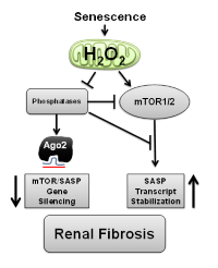
Donald McCarthy: 2014, “Redox-control of the senescence associated secretory phenotype”, SUNY at Albany, CNSE,
Current Position: Post-Doctoral Fellow, UCSF

Anthony Franchini: 2014, “Redox-Regulation FcR-receptor signaling and macrophage function”, Albany Medical College.
Current Postion, Post-Doctoral Fellow, University of Rochester Medical Center.

Nicole Flaherty: 2016, “Control of F. Tularensis pathogenesis through TRPM2”

A. Chandrasekaran: 2017, “Redox-control of TRPC6 and the senescence associates secretory phenoytpe”


Master Thesis Committees

Raichal Melathe: 1998, “Elucidation of transcript differences in superoxide dismutate 2 overexpressing tumor cells”, Albany Medical College
Current Position: Laboratory Manager Montefiore Medical Center.

Kwi-hye Kim: 2003, “Redox-dependent control of Nitration through an altered antioxidant enzyme status”, Albany Medical College
Current Position: Postdoctoral Scientist at Eli Lilly and Company

Supriya Kar: 2010, “In vivo antioxidant deficiency augments age-associated MMP-1 expression” Albany Medical College
Current Position: Pediatrician, Hartford CT

Aldo Gammara: 2006, “Redox-control of TIMP expression”, Albany Medical College
Current Position: General Surgery private practice Staten Island, NY.

Lisa Keenan: 2014, “The role proly hydroxylase in the regulation of MMP expression”, SUNY Albany, CNSE

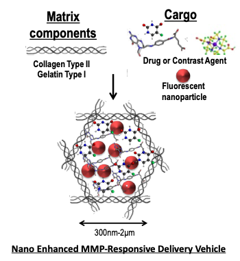
Ahmad Nazem: 2016, “Characterization of Degradome Sensing Nanoparticles for treatment of degenerative Disease”, SUNY Albany CNSE

N. Nerlakanti: 2016, “Development of Protease Sensing surface plasmon resonance array”, SUNY Albany CNSE

Y Lee: Current, “Epitranscriptomic control of selenocysteine utilization and senescense”


Former Post-Doctoral Associates

Nadine Hempel
Associate Professor, Penn State Medical College, Hershey, PA
Assistant Professor, College of Nanoscale Science & Engineering, University at Albany, 2011-15
Postdoctoral fellow, 2007-2011

Toni Bartling
Staff Scientist, GE Health Sciences
Postdoctoral fellow, 2009-2013

Hanqing Ye
Research Associate Hunan University, China
Postdoctoral fellow, 2006-2009

Pauline M. Carrico
Instructor, Biology, University at Albany, 2013-present
Assistant Professor (non-tenure track), 2000-2003, Assistant Professor Empire State College, Saratoga, NY.
Postdoctoral fellow, 1999-2000


Former Undergraduate Students

Name Year of Graduation University
Faith Williams 1998 SUNY Albany
Sian Martin 1998 SUNY Albany
Lisa Schoonmaker 1999 Siena
Kevin Regan 2000 SUNY Albany
Martin King 2001 University of Illinois
Cynthia Bernal 2005 SUNY Albany
Bharat Yarlagadda 2005 Rensselaer Polytechnic Institute
Young In Lee 2005 Yale University
Chidiebere Nwaogu 2006 Albany Medical College
Harlan Rozenberg 2007 SUNY Albany
Bryan Abessi 2008 Lemoyne
James Joseph 2009 SUNY Albany
Jocelyn Laboy 2009 SUNY Albany
Katie Wang 2009 U. of Rochester
Lina Bagepali 2010 Rensselaer Polytechnic Institute
Michael Taylor 2010 Georgetown University
Vivek Bhatty 2010 SUNY Albany
Nilay Patel 2011 Rensselaer Polytechnic Institute
Marcelle Tuttle 2012 Boston University
Eric Kelley 2012 SUNY Albany
Brooke Pati 2012 SUNY Albany
James McNeilan 2012 SUNY Albany-CNSE
Teresa Regis 2013 SUNY Albany
May Lee 2015 SUNY Polytechnic-CNSE
Jeff Richards 2015 SUNY Polytechnic-CNSE
Leo Bezerra 2015 SUNY Polytechnic-CNSE
Tristin Schwartze 2016 SUNY Polytechnic-CNSE
Max Matiaude 2017 SUNYPolytechnic-CNSE
Russel Shapiro 2017 SUNY Polytechnic-CNSE

Facilities

Melendez

The PI occupies a laboratory of approximately 1400 sq. ft. of floor space and 200 linear feet of bench. The PI’s lab is equipped with a sinks, chemical and tissue culture hoods, dishwasher, autoclave, microplate reader, ABI 7500 RT thermal cycler, Eppendorf mastercycler, water baths, rotary shakers, micro-centrifuges, a refrigerator/freezer, -80 freezers, UV/VIS spectrophotomoter and gel documentation system. The lab is also equipped with 3 laptop computers for data analysis and manuscript preparation. The PL’s tissue culture room contains two 6 ft laminar flow hoods, 4 Forma steri-cult incubator, 1 Forma low-gas steri-cult incubator and Nikon Diaphot 200 inverted microscope including bright field and epi-fluorescence capabilities, dichroic cube sets B-2E and G1-B, Nikon objective lens set including: CF Fluor 20x, O.75 n.a., CFN Plan Fluor 40x, 0.7 n.a., CFN 60x oil Plan Apochromat, 1.21 n.a., CFN 100x oil Plan Apochromat, 1.4 n.a. lenses.inverted microscope. The laboratory also has 2 Mettler top-loading balances, Savant Speed-Vac concentrator, Beckman dual-beam spectrophotometer, Biotek Synergy UV/VIS/Fluor/Chemiluminescence plate reader Gibco/BRL horizontal agarose gel units and polyacrylamide slab gel units, 10 LKB power supplies, Forma -80C freezer, 3 Freezers and 2 refrigerator/freezer, 2 Shaking incubators, 2 bacterial/yeast incubators, 2 Chemical hoods. Amnis ImageStream (Amnis Corporation) imaging flow cytometer that is equipped with 488 nm and an extended depth of field module. This instrument, which operates in a Windows-based environment, introduces a new technology platform that captures high-resolution digital images of cells in flow at rates of approximately 100 cells/per second. It simultaneously acquires up to six different images of each cell, including four colors of sensitive fluorescence imagery, side scatter and brightfield imagery. The ImageStream combines the quantification and analysis of cellular morphology with the ability to perform all the fluorescence intensity measurements of conventional flow cytometry, allowing for rapid analysis and classification of thousands of cells using the ImageStream IDEAS software. The IDEAS software calculates over 100 parameters per cell, including all the standard intensity-based parameters and statistics employed in flow cytometry as well as numerous morphological parameters such as cell area, perimeter, aspect ratio, texture, spot counts and internalization ratios. The ImageStream is ideally suited for quantitative investigations of nuclear translocation, phagocytosis, intracellular co-localization, apoptosis, cell: cell interactions and fluorescent in situ hybridization in suspension. Meso Scale Discovery SECTORTM Imager 2400 (SI2400) instrument that enables either a single or multiple analytes/biomarkers to be determined simultaneously within a single well using a small amount of sample. It has exceptional sensitivity, wide dynamic range and convenience using a line of custom microplates. The SI2400 instrument accommodates 96- and 384-well single-spot plates as well as 96-well multi-spot plates. This platform is ideal for large scale clinical studies and screening.The PI also has full access to a Zeiss AxioVert200M fully automated inverted microscope with motorized stage quipped for multicolour, multiposition live cell imaging and feature .live cell, phase Contrast, bright field, DIC, color camera, Z-stack and timelapse.

Computers

Multiple Lenovo desktop computers with internet access are available, all equipped with appropriate software for data analysis, word processing, email, etc. Computers and infrastructure are supported by CNSE computer department. The PI and his students also have access to OriginLab, Statistica and GraphPad Prism (plotting and statistical analysis software); ANSYS Multiphysics, FLUENT, IntelliSuite and COMSOL (finite element analysis and simulation software); as well as L-Edit, Cadence, and K-Layout (lithographic layout software) that is hosted on the CNSE network and is supported by the CNSE Supercomputer Core Facilities.

Shared equipment: includes -150°C Revco, -80°C and -20°C freezers, +4°C refrigerators, acid fume hoods, four Thermo Scientific CO2 incubation chambers, four Class II/A2 biological safety cabinets,Tecan Infinite M200 microplate fluorometer with UV/VIS spectrophotometer capability, BioRad Protean II gel electrophoresis equipment, BioRad VersaDoc 4000 Imaging System, NanoDrop ND-1000, Spectrophotometer, NanoDrop ND-3300 Fluorospectrometer, Malvern Zetasizer Nano, centrifuges, autoclave, and a humidity controlled BioForce Nano eNabler (NeN) QPL instrument (Bioforce Nanosciences) for cell printing adjacent to cell incubators and fluorescent microscopes.

CNSE Microscopy and Analytical

The CNSE complex also has a full suite of metrology tools including electron and ion microscopes (TEM/SEM/FIB), light microscopes, X-ray photoelectron spectroscopy, atomic force microscopes and other surface analysis and microscopy tools. Also available are a Bruker D8 ADVANCE X-ray Diffractometer (HR-XRD), Thermo VG Scientific Theta Probe X-ray Photoelectron Spectroscopy (XPS), Digital Instruments Nanoscope III Scanning Probe Microscope (SPM/AFM) Multimode and 3100 system with liquid cell, LEO 1550 Scanning Electron Microscope with a ThermoNoran Voyager Energy Dispersive X-ray Detector and a MaxRAY WDS system (SEM), JEOL 2010F High Resolution Transmission Electron Microscope (TEM) with EELS and a Nanofactory TEM-STEM holder, FEI Nova NanoLab 600 Dual Beam Focused Ion Beam/Scanning Electron Microscopy System (FIB/SEM), Nicolet 6700 FT-IR with transmission and diffuse reflection measurement capability. A J.A. Woollam InfraRed-Variable Angle Spectroscopic Ellipsometer (IR-VASE) is available for transmission and reflection measurements on thin films and monolayer chemistries. ICP: Perkin Elmer Nexion 300 ICP-MS, The Nexion 300 is a bench top ICP-MS (Inductively Coupled Plasma Mass Spectrometer) with a quadrupole mass analyzer that is capable of performing trace elemental analysis. The instrument was recently installed and will be an open access instrument. Users are granted access after receiving training.

Additional Resources available at UAlbany-SUNY Life Science Research Building Core Facilities

Molecular Core Facility: includes a real-time PCR machine (ABI 7900HT), several traditional PCR machines (ABI), a low volume spectrophotometer (NanoDrop), centrifugues (from desktop models to high-speed ultracentrifuges), BioRad ChemiDox XRS, GE Typhoon Trio phosphoimager, BioRad Pathfinder FPLC, PCR/DNA sequencing, bacterial shakers, and basic molecular biology equipment and microscopes: MMI LCM microscope, Leica fluorescent dissecting microscope, Nikon TS100 microscope. Biological Imaging Facility: Includes a Zeiss 940 Digital SEM, Zeiss 902 TEM, Zeiss LSM510/META scanning head confocal microscope, and Zeiss AxioVert 100M inverted fluorescence microscope with 20X Plan ApoChromat (NA 0.75, WD 0.61), 40X AchroPlan (water, NA 0.8, WD 3.61 mm), 63X PlanApochromat (oil, NA 1.4, WD 0.18) and 100X PlanNeofluor (oil, 1.3, WD 0.17). Digital imaging includes Metamorph and NIH/Scion software; Hammamatsu ORCA CCD cameras, and high quality calcium-sensitive fluorescence imaging via Metafluor. Photography facilities are also present. Tissue Culture Core Facility: The tissue culture facility is a biosafety level 2 facility and contains basic tissue culture hoods, water-jacketed incubators, a liquid nitrogen cell freezer, and -80C freezers. Crystallography Facility: Bruker D8 Advance X-Ray powder diffractometer equipped with a Globel mirror and a Super Speed Linxeye positional detector, Bruker CCD Single Crystal X-Ray diffractometer equipped with a cooling device. Structural Chemistry Core: Fluorolog-3 fluorescence spectrometer, Lambda-950 UV/Vis/Near IR absorption spectrophotometer, Spectrum-100 FT-IR spectrometer, JASCO-400 stopped-flow instrument, Raman Microscope.

Support

Current Support

  • CNSE

Past Support

  • NIH
  • SEMATECH/ Nano Health & Safety Center (CNSE / SUNY PI)
  • Phillip Morris