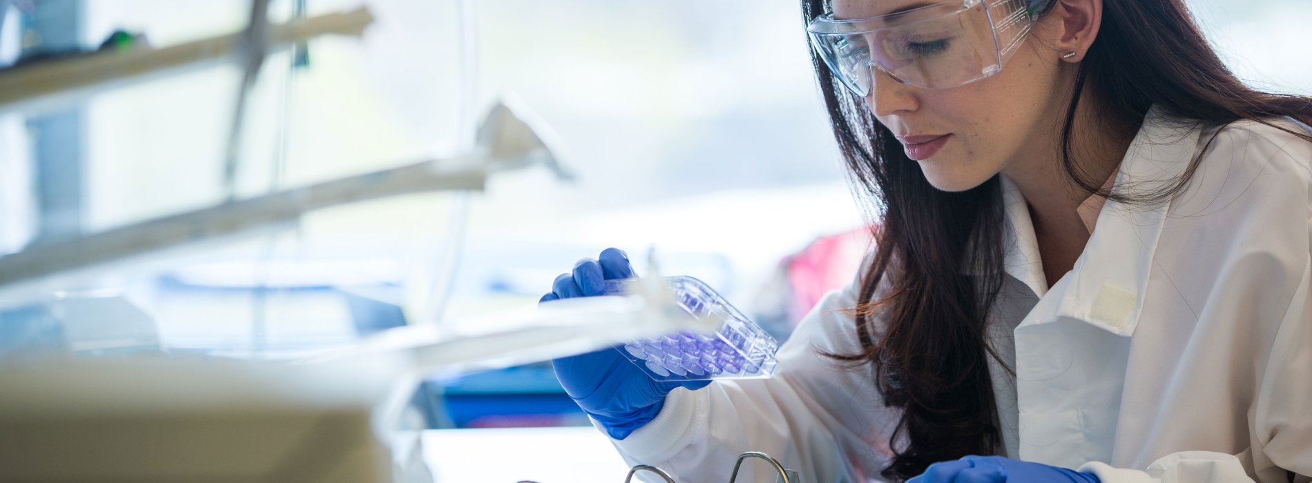
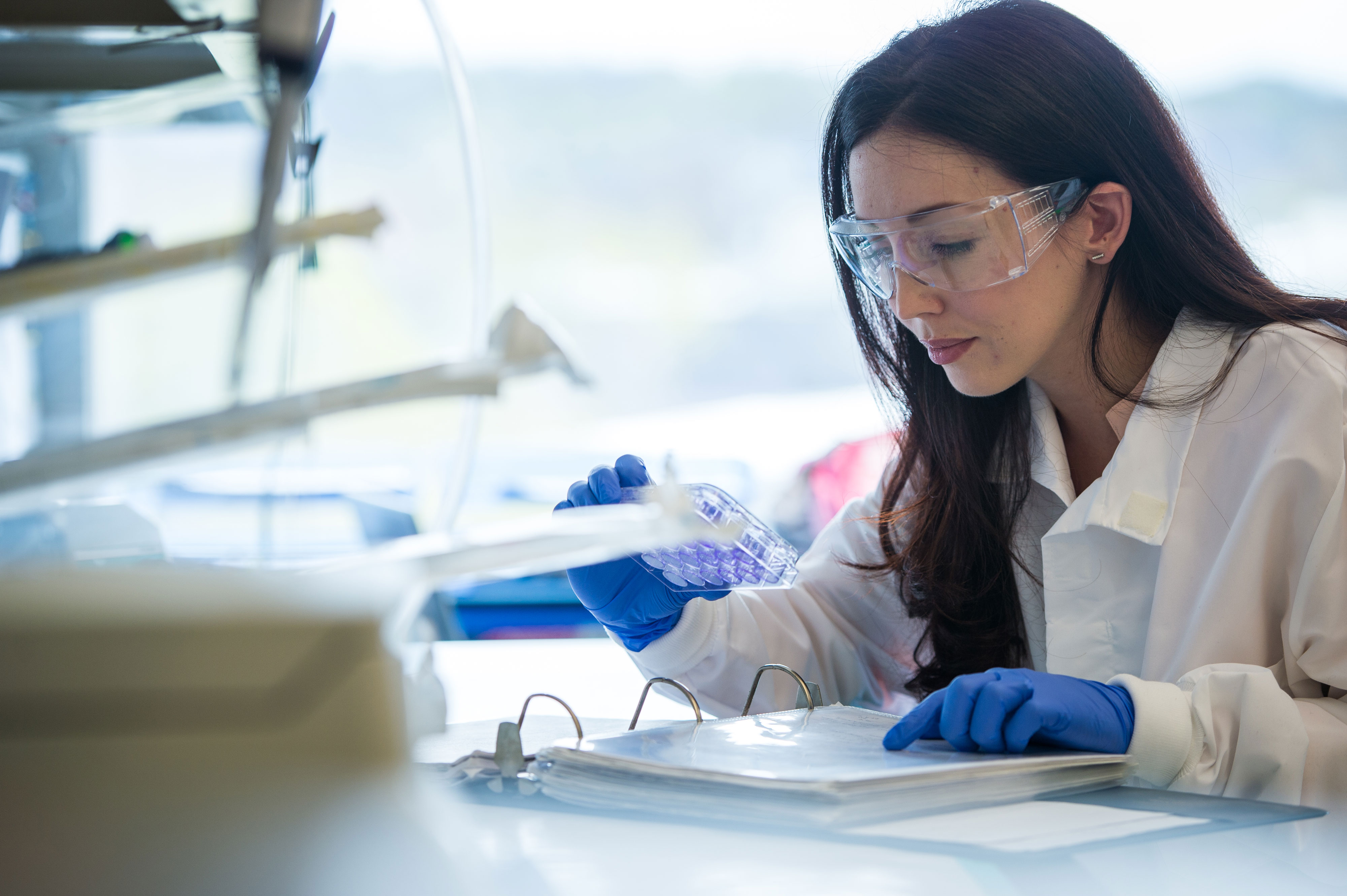


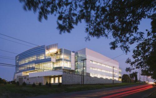
Researchers at the UAlbany Cancer Research Center are working to better understand the genetic and environmental causes of cancer, while providing in-depth training to early-career scientists.
Using state-of-the-art technologies, faculty and students focus on the underlying biology associated with tumor initiation and progression, as well as the development and evaluation of chemo-preventive regimens and therapeutic approaches for common cancers.
Our dedication to advancing cancer research while educating the next generation of scientists is demonstrated by various awards, including a $1.7M grant from the National Cancer Institute to study the role of nutrition in breast cancer.
You can support our efforts to eliminate cancer by contributing to the Fund for Memory and Hope. Gifts of any amount are most welcome.
Donate Today Attend an Event Meet our Researchers
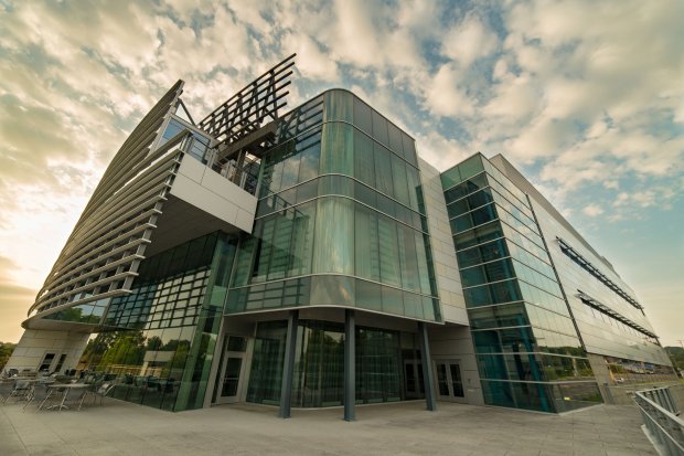
The Cancer Research Center is housed within UAlbany’s School of Public Health, which was founded in partnership with the New York State Department of Health — a close connection that makes the Center unique among cancer research facilities in the U.S.
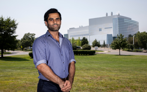
Our faculty provide a collaborative research environment to foster graduate students and postdoctoral fellows' training in cancer biology.
Explore our master's and doctoral programs in Biomedical Sciences and Environmental Health Sciences.
Faculty at the Cancer Research Center address a variety of topics related to cancer, including:
Understanding the molecular actions of natural products including vitamins and dietary components
Defining the genomic and molecular events associated with progression of early-stage disease to aggressive cancer
Exploring whether exposure to silver nanoparticles — which can be found in a wide variety of household products—can lead to the development of cancer
Identifying and understanding the genetic susceptibilities of cancer cells
Investigating Myalgic Encephalomyelitis (ME) / Chronic Fatigue Syndrome (CFS) to explore genetic and immunologic links between ME/CFS, autoimmune disorders and cancer

Cancer Research Center Journal Club: Campus community members gather to watch a student, postdoc or faculty member present a paper of interest. Join us for a lively scientific discussion at 11 a.m. Tuesdays in the Cancer Research Center’s Massry Conference Room.
Friday Research Talks: Join students, faculty and staff members for networking and informal conversations about ongoing research projects. These discussions are held at 11 a.m. Fridays in the Cancer Research Center’s Massry Conference Room.
Established through the generosity of Daniel J. Hogarty and the Hogarty Family Foundation, the annual Hogarty Lecture at the Center Research Center provides information to the community about a broad range of topics related to cancer.
Experts have previously spoken on cancer control in the 21st century, aging and cancer, inflammation and cancer progression, and combatting the tobacco epidemic.

1 Discovery Drive
Rensselaer, NY 12144
United States